Lower Limb Muscles and Ligaments: What Are the Key Structures and Their Functions?
Published on 02/25/2025 · 11 min readUnderstanding the intricate network of muscles and ligaments in your lower limbs is fundamental to comprehending movement, stability, and potential injuries. Let's delve into the key structures of the hip, thigh, and gluteal region.
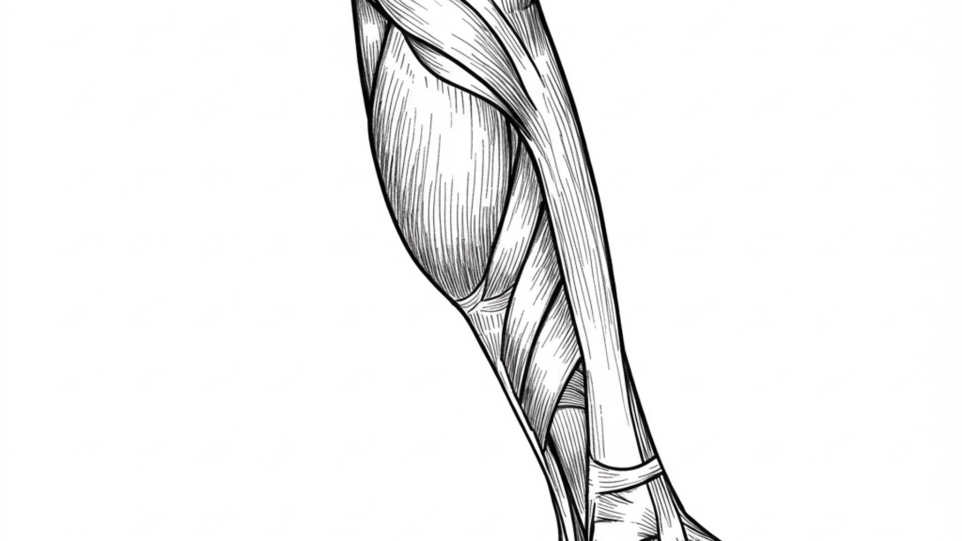
Table of Contents
Key Ligaments of the Lower LimbMuscles of the Gluteal Region (Your Butt)Muscles of the ThighAnterior Compartment (Femoral Nerve)Medial Compartment (Obturator Nerve)Posterior Compartment (Sciatic Nerve)
Key Ligaments of the Lower Limb
Ligaments provide crucial stability to joints. Here are some important ones:
- Inguinal Ligament: Extending from the anterior superior iliac spine to the pubic tubercle, it's the lower border of the external abdominal oblique aponeurosis. The femoral artery passes under it.
- Ligament of Cooper: Found in the pelvis and also in the breast, providing structural support.
- Obturator Membrane: Mostly closes the obturator foramen in the hip bone, with the obturator canal allowing passage of the obturator nerve.
-
Sacrospinous Ligament: Runs from the sacrum to the ischial spine, dividing the sciatic foramen into greater and lesser parts.
- Greater Sciatic Foramen: Transmits the piriformis muscle, superior and inferior gluteal vessels and nerves, sciatic nerve, posterior femoral cutaneous nerve, nerve to obturator internus, nerve to quadratus femoris, internal pudendal vessels, and pudendal nerve.
- Lesser Sciatic Foramen: Transmits the tendon of obturator internus, nerve to obturator internus, internal pudendal vessels, and pudendal nerve.
- Sacrotuberous Ligament: Connects the lateral sacrum and posterior iliac crest to the ischial tuberosity.
Muscles of the Gluteal Region (Your Butt)
These muscles are vital for hip movement and stability.
- Tensor Fasciae Latae: Originates from the iliac crest and inserts into the iliotibial tract. Functions to tense the fascia latae and abduct the hip.
-
Gluteal Muscles:
- Gluteus Maximus: The largest muscle, originating from the ilium, sacrum, and sacrotuberous ligament, inserting on the gluteal tuberosity of the femur and iliotibial tract. Responsible for hip abduction, extension, and lateral rotation.
- Gluteus Medius: Originates from the ilium and inserts on the greater trochanter. Functions in hip abduction and medial rotation, crucial for preventing pelvic tilting (Trendelenburg gait). Supplied by the superior gluteal nerve.
- Gluteus Minimus: Deep to gluteus medius, with similar origin and insertion, also contributing to hip abduction and medial rotation and preventing pelvic tilting. Supplied by the superior gluteal nerve.
-
Lateral Rotators of the Hip (External Rotators):
- Piriformis: Originates from the sacrum and passes through the greater sciatic foramen, inserting on the greater trochanter. Nerve supply from S1 and S2.
- Quadratus Femoris: From the ischial tuberosity to the femur. Nerve to quadratus femoris.
- Obturator Internus: Originates from the inner obturator membrane, tendon passes through the lesser sciatic foramen. Nerve to obturator internus.
- Obturator Externus: Originates from the outer obturator membrane and inserts on the femur. Supplied by the obturator nerve.
- Superior Gemellus: Superior to obturator internus. Nerve to obturator internus.
- Inferior Gemellus: Inferior to obturator internus. Nerve to quadratus femoris.
Muscles of the Thigh
The thigh is divided into anterior, medial, and posterior compartments.
Anterior Compartment (Femoral Nerve)
- Quadriceps Femoris: A group of four muscles (rectus femoris, vastus medialis, vastus lateralis, vastus intermedius) with a common insertion on the patella and tibial tuberosity via the patellar ligament. Responsible for hip flexion and knee extension. Note: Osgood-Schlatter disease affects the tibial tuberosity.
- Sartorius: The longest muscle, from the anterior superior iliac spine to the medial tibia. Aids in hip flexion, abduction, and external rotation, as well as knee flexion.
- Iliopsoas: A combination of psoas major (lumbar vertebrae) and iliacus (iliac fossa) muscles, inserting on the lesser trochanter of the femur. Powerful hip flexor and also involved in medial rotation of the thigh and flexion of the pelvis on the thigh (sit-ups).
Medial Compartment (Obturator Nerve)
These muscles primarily adduct the hip.
- Gracilis: From the pubis to the upper medial tibia.
- Pectineus: From the pectineal line of the hip bone to the upper femur.
- Adductor Longus: From the pubis to the linea aspera of the femur.
- Adductor Brevis: From the pubis to the linea aspera of the femur.
- Adductor Magnus: A large muscle originating from the ischial tuberosity and ischiopubic ramus, with insertions on the linea aspera and adductor tubercle of the femur. The adductor hiatus allows passage of femoral vessels to become popliteal vessels.
Posterior Compartment (Sciatic Nerve)
These muscles extend the hip and flex the knee.
- Biceps Femoris: Two heads (long and short) originating from the ischial tuberosity and linea aspera, inserting on the fibular head.
- Semitendinosus: From the ischial tuberosity to the medial tibia.
- Semimembranosus: From the ischial tuberosity to the medial tibial condyle. Note: These muscles border the popliteal fossa.
This overview provides a foundational understanding of the major muscles and ligaments of the lower limb. For a more in-depth study, consult detailed anatomical resources.
Shop related blood tests
This genetic marker is associated with certain inflammatory and autoimmune conditions, some of which can affect the joints and surrounding tissues of the lower limbs (e.g., ankylosing spondylitis). While the lecture is primarily anatomical, recognizing potential underlying inflammatory conditions that could impact these structures is clinically relevant.
Read next
 Written on 02/01/2025
How Does Erythropoiesis Work? Understanding Red Blood Cell Formation, Iron, B12, and Folate's Role in Anemia and Polycythemia.
Erythropoiesis, the process of red blood cell (RBC) formation, is essential for maintaining oxygen transport throughout the body. This intricate process involves multiple factors, including erythropoietin (EPO), iron, vitamin B12, and folate. Understanding these components is crucial for comprehending conditions like anemia and polycythemia. Read more
 Written on 03/06/2025
What Are the Core Tenets of Cell Theory, and How Do Prokaryotic and Eukaryotic Cells Differ?
Let's break down the word "biology." As the study of etymology tells us, "bio" means life, and "logy" (from "logos" or "logia") means the study of. So, simply put, biology is the study of life. And what's the basic unit of life? You guessed it – the cell! Our bodies are made up of around 100 trillion cells – pretty mind-blowing, right? Read more
 Written on 02/22/2025
What Is Tinnitus? Causes and Treatment
Tinnitus is a perception of noise or ringing in the ears or head when there is no external source of sound. Read more
 Written on 03/07/2025
What Are Centromeres and Telomeres? Exploring Their Structure, Function, and Role in DNA Replication and Cell Aging.
Ever wondered how our genetic material, DNA, is organized and protected within our cells? Two key structures play vital roles in this process: centromeres and telomeres. Let's dive into their fascinating world and understand their significance in cell division and the aging process. Read more
